
Clinics
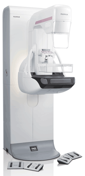
Aspire Cristalle
NEW IN CANADA, this Fuji mammography system offers SMARTER patient comfort, departmental workflow, and image quality.
Built to perform, ASPIRE Cristalle, digital breast tomosynthesis offers a patented comfort design for a better patient experience, a superior diagnostic accuracy for radiologists, and fast, easy to use, reliable workflow.
Fujifilm’s ASPIRE Cristalle includes:
Super diagnostic accuracy through:
- Adaptative image processing. To produce consistent image quality across the wide range of patients, ASPIRE Cristalle incorporates image processing algorithms that automatically “adapt” to patients breast thickness and composition. Using analytical processes similar to those used with iAEC, these algorithms localize the glandular regions in the acquired image, assess their contrast, and if needed, make minor adjustments to default processing parameters.
- Iterative Super-Resolution (ISR) reconstruction generates initial slice images; simulated projection images from the slice images; and compares the simulated projection images to the actual projection images. ISR also calculates the amount of error and generates in slice images to decrease it.
Streamlined workflow through fast image acquisition and display; twice as fast, with a 15 seconds 2D cycle time.
Advance in dose reduction by approximately 20% through:
- Image based Spectrum Conversion (ISC) By performing an analysis of the energy distributions between the two types of target materials, Fujifilm has developed an image processing algorithm that takes advantage of W target’s lowest dose imaging, while, at the same time, providing the high contrast images of Mo. ASPIRE Cristalle introduces this new feature.
- Hexagonal Close Pattern Detector. By utilizing innovative hexagonal pixels, the electric field between them is considerably stronger and they improve the information capture.
- Intelligent Automatic Exposure Control that performs a detailed analysis of the pre-exposure image to determine the optimum exposure conditions for the view.
- Three dose modes (H, N, andL) that can be selected by the technologist (Nmode is typically used). These modes coupled with the increased sensitivity of its HCP detector and improved dose stabilization of its iAEC method, enable ASPIRE Cristalle to demonstrate reduced patient dose when compared to a competitive FFDM system.
Increased diagnostic confidence through:
- Dynamic Vision II locates pectoral muscle and implants to exclude them from further analysis. It also identifies glandular and adipose tissues, and features. DVII applies MFP2 processing for edge enhancement & dynamic range control, and offers better conspicuity of microcalcifications, improved visibility and morphology of masses compared to conventional processing due to 100 mm pixel output, lower exposure dose to patients and DVII image processing.
- Tomosynthesis has an X-ray tube that moves through an arc, acquiring a series of low-dose images at different angles, and then, reconstruct them into a series of high-resolution slices which are 1 millimeter apart. This reconstruction makes it easier to identify lesions.
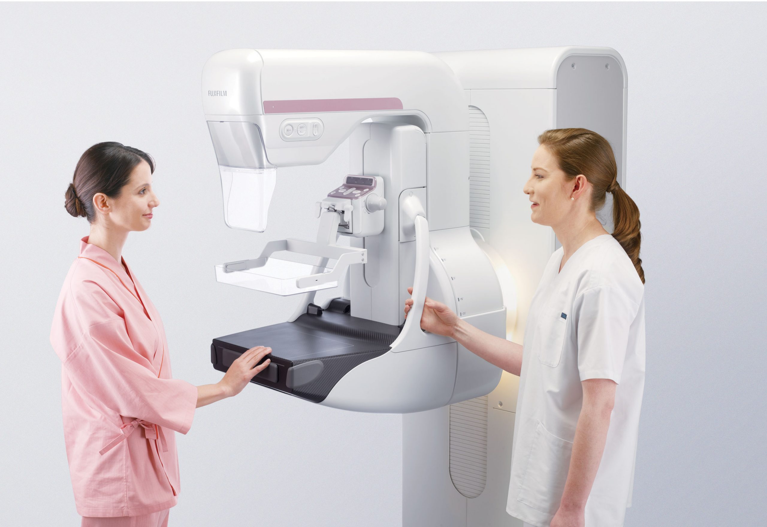
A more comfortable exam through:
- Comfort paddle gently adapts to the breast, allows for firmer, more tolerable compression and plate to flex and contour to the breast. It also distributes pressure evenly, for more even distribution of breast tissue.
Autoaligned Technology
Reliability & dependability through one shot quality control program that provides all phantom materials and software support needed for QC Technologist and Medical Physicist testing; and allows for trending and print out of accumulated weekly, quarterly, semiannual & annual QC test results.
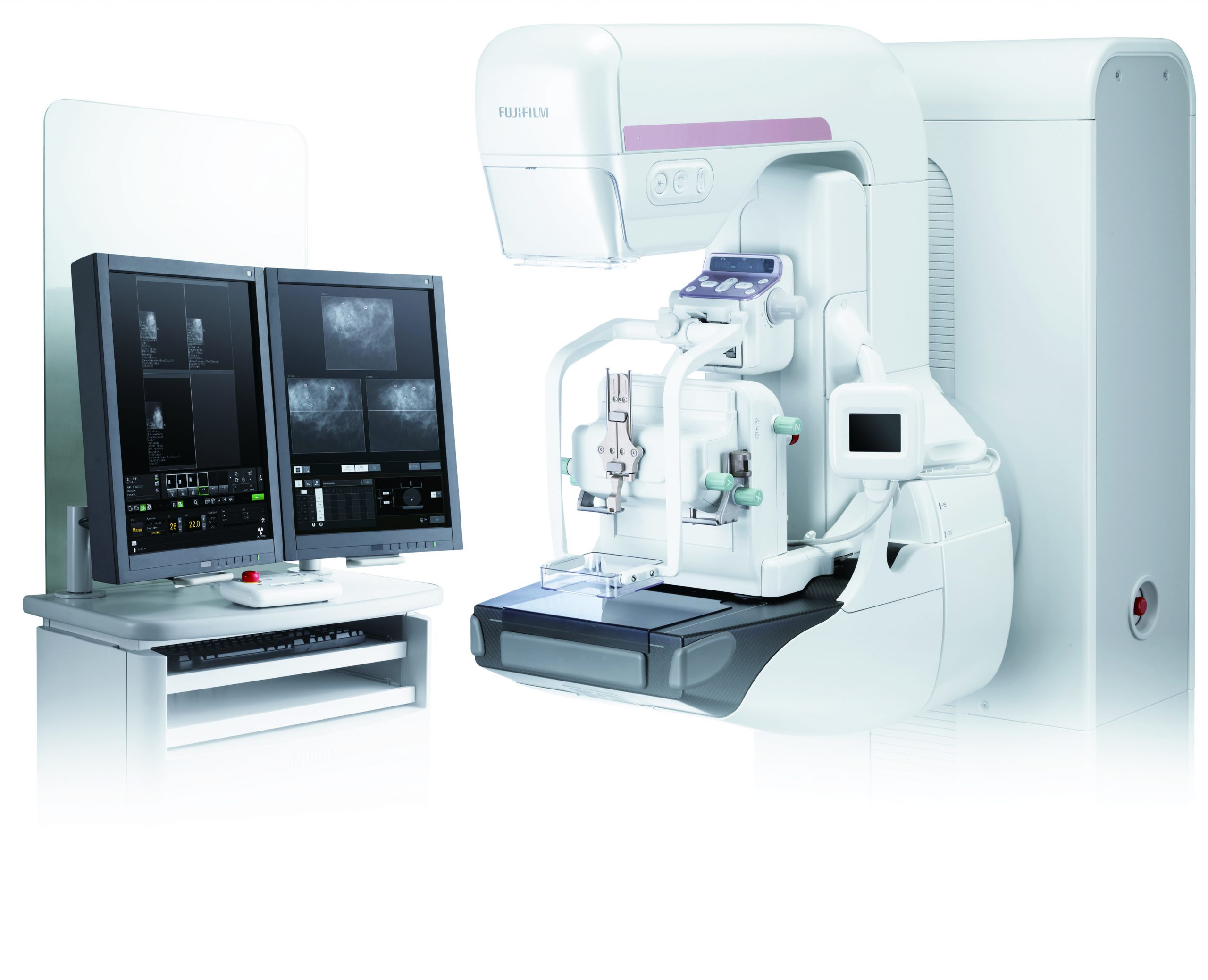
Stereotactic biopsy
Our Stereotactic Positioner delivers simplified needle placement and visualization for improved diagnostic confidence.
- Easy to use for the radiologist and the technologist
- Users can manipulate and enhance the image in real time
- Multi-vendor needle compatibility for VAB, CNB and Hookwire
- Tube and detectors are flexible from—90° to +90°, offering more positioning options
- Patient positioning for stereo biopsy either upright or recumbent
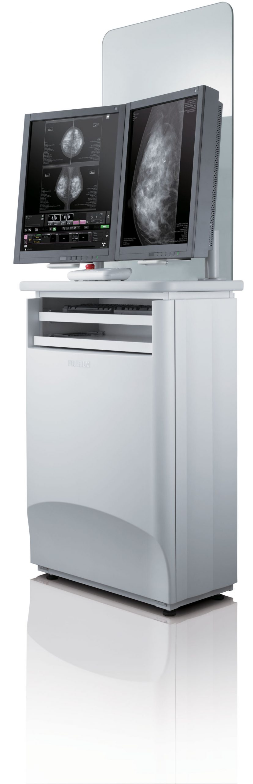
The attributes that make DBT so valuable to your clinical practice cause pressure on your IT. The enormous datasets can cause significant study management challenges, making it imperative that you consider your IT strategy carefully.
Our Synapse® Enterprise Imaging portfolio – including Synapse 5 PACS and Synapse VNA – enables secure, easy-to-manage storage and access to the complete patient imaging record, and this, throughout the healthcare enterprise. Our Synapse 5 server-side rendering technology is ideally suited to handle the massive datasets generated by DBT exams—enabling unprecedented image-rendering speed, a reduced burden on IT infrastructure and increased speed to diagnosis. It immediately outputs individual images to PACS, viewer or printer during exam , improves system availability and extends system lifespan through programmable automatic startup, sleep and shutdown.
Built smarter for image quality
The detector is the heart of any digital mammography system. Simply put, its job is to detect X-rays, convert them into electrons, and collect the resulting electrical charges. The more efficiently it collects charges, the stronger the image signal, the less noisy the image, and the lower the dose needed.
Hexagonal Close Pattern (HCP) pixel design
In conventional detector design, the pixels that detect X-rays are square, with wide gaps between them, losing some of the converted X-ray information.
In the ASPIRE Cristalle, hexagonal pixels are arranged with smaller gaps between pixels, resulting in less signal loss, stronger electrical fields, and higher sensitivity. When compared to square pixels, HCP delivers:
- 20% increase in detector sensitivity
- Improved information capture
- Lower patient dose
Optimized Dose for Maximum Image Quality
ASPIRE Cristalle is built with Intelligent Automatic Exposure Control (iAEC). iAEC intelligently analyzes breast composition and thickness and applies auto-recognition of implants. This optimizes dose and processing to generate exceptional images at low doses for all breast types, including breasts with implants.
![]()
One vendor, one solution
From DR and CR digital X-ray systems, to women’s health imaging systems, to the Synapse family of products that includes PACS, RIS, Synapse 3D, and Synapse Cardiovascular, to archiving and cloud storage, Fujifilm provides a comprehensive portfolio of solutions that allows health systems to integrate and consolidate data management, standardizing platforms across all imaging. Fujifilm provides the answers to increasing pressure on the efficiency and productivity of technologists and radiologists.
Fujifilm: Focused on Continuous Innovation
Fujifilm Medical Systems is devoted to providing healthcare experiences that enhance the quality of life. From the development of the first web-based PACS system to new detector technologies such as ISS and Direct Optical Switching, Fujifilm Medical Systems continues to provide technological solutions that embody these values.
Fujifilm is constantly pursuing better solutions, from microscopy to outer space. Fujifilm Advanced Research Laboratories works in fields ranging from organic synthesis, to pharmaceuticals, to image processing, bringing continuous innovation and leading-edge products to a broad spectrum of industries. Fujifilm is a Fortune 200 global corporation powered by a dedicated international workforce, an annual commitment of $2 billion to research and development (R&D), and a steadfast pledge to empower customers through innovation. Fujifilm is dedicated to researching and developing cutting-edge technologies that will shape the future.