Clinics
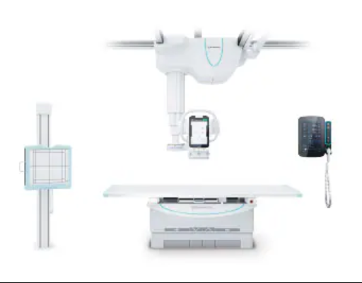
RADspeed Pro SR5 Version
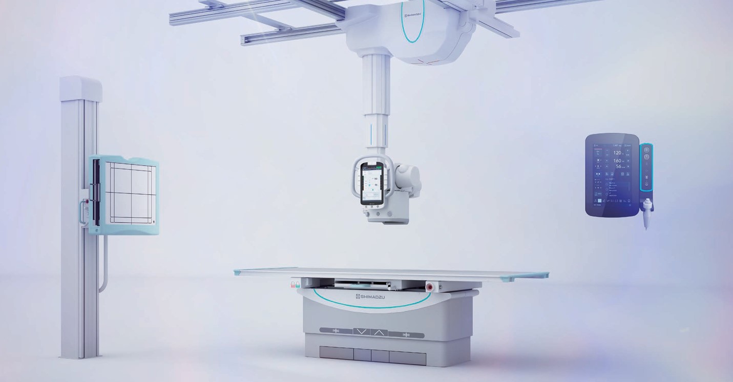
The high-performance general radiography system allows to increase examination efficiency by standing closer to patients and reducing the burden on medical personnel.
Vision for Easy Operation
Healthcare requires multiple complex tasks. To support this hectic work, it is essential to achieve an examination environment that contributes to diagnosing patients, while also ensuring simple and intuitive operability. Shimadzu offers systems optimized for usability.
Optical camera apps increase examination efficiency. Enhanced imaging operations reduce the burden on medical personnel.

This system is based on the extensive experience and expertise Shimadzu cultivated as a pioneer in medical diagnostic imaging systems. It minimizes X-ray dose and improves examination workflow. Available only from Shimadzu, this system utilizes the latest technologies that are gentler to patients and provide a more comfortable examination environment for the patient and operator alike.
- Abdominal/Contrast
- Musculoskeletal
- Pediatric
- Scoliosis
- Leg length
- Pulmonary CXR
- Optical Camera Application Creates an Environment where Medical Personnel can Focus on Patients
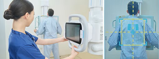
The video image from a camera built into the collimator is displayed on the X-ray tube support control panel and high-voltage generator control panel monitors. The optical camera application provides an environment where medical personnel can focus on patient care.
- Live View Display: Reduces Positioning Effort and Improves Accuracy
Supports accurate positioning by showing overlay of detector area, irradiation field and AEC pickup fields*, which are difficult to check directly.

- Motion Detection: Reduces Frequency of Repeating Exposures due to Body Movement
Patient body movement can be confirmed from the point that body movement detection mode is activated.*

*Check the patient’s condition, even directly visually.
- Last Position Display: Smoother Positioning Correction during Repeated Exposures
By checking the immediately previous exposure positioning, positioning can be achieved more smoothly when repeating exposures.

- Illumination Improves Visibility
Illumination of the X-ray high-voltage generator and ceiling-mounted X-ray tube support enables better understanding of the instrument status. In addition, the hand switch illuminates to indicate the system is ready for the next exposure.

- Lower Hand Grip: Easy to operate in high positions
A hand grip is provided on the back side at the bottom of the control panel, and operation is possible by pressing the all-free switch on the front side. Operation is easy even when the X-ray tube support is located in a high position.

- Intuitive operation: Graphic Display of Unlock Buttons
Graphic unlock buttons enable more intuitive operability by displaying button symbols with the unlock direction oriented to match the perspective of the operator in either supine or standing positions.

Assist Functions Support Positioning
- Superb Operability: Vision for Reduction of operator burden
A wide range of positioning support functions reduces the operator burden.
![]()
Motors assist handle operations. This reduces the burden on operators during movements by enabling the ceiling-mounted X-ray tube support to be moved quickly and lightly.

High Throughput: Vision for High Throughput
Use it to perform examinations smoothly, while relieving patient anxiety. To achieve both, a system is required that can shorten examination times while ensuring safety. Shimadzu supports efficient examination process flows for front-line healthcare workplaces.
-
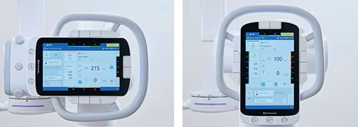
- Enhanced Visibility: Equipped with a larger screen on the X-ray tube, making it easier for technologists to view and capture images with confidence.
- Upgraded Generator Console: A redesigned console for seamless, intuitive control.
- Effortless Handling: Improved ergonomic design for easier handling and mobility in any healthcare environment.
- Integrated Camera with AI Motion Detection: Advanced AI-powered motion detection tools for improved automation and accuracy.
- Enhanced Cable Management: Tidy, efficient cable management to streamline the entire workspace.
- Power Glide Motorized Assistance: Motorized assistance to support technologists, reduce strain, and enhance overall operational ease.
- Live View Display: Supports accurate positioning by showing overlay of detector area, irradiation field and AEC pickup fields, which are difficult to check directly.

To carry out our role in the early diagnosis of disease and the improvement of cure rates, Shimadzu provides a broad range of diagnostic imaging equipment.
At this time, a number of innovations are occurring at the leading edges of medical treatment. In the field of diagnostic imaging, Shimadzu has developed the direct conversion flat panel detector (FPD) that provides heretofore unavailable high-quality images. We are also the first in the world to market the circulatory organ diagnostic system on which this detector is mounted. Additionally, we have developed a variety of other diagnostic systems that utilize this FPD. Shimadzu is now a leading pioneer of these new types of diagnostic imaging.
Furthermore, recent IT technology developments are introducing efficient diagnostic systems to a variety of medical treatment areas. Shimadzu is supporting the renovation of IT systems in hospitals and medical centers by providing digital processing systems that incorporate the newest IT technology wherever possible and can process many kinds of medical information, including examination images.
Request a demo today!
Our innovative Medical Imaging Equipment Solutions and Health IT Solutions are built to support your practice with the latest advancements in medical imaging. Experience how our solutions can enhance diagnostic accuracy, streamline workflows, and improve patient care. Click to request a demo and discover the possibilities firsthand.