

Clinics
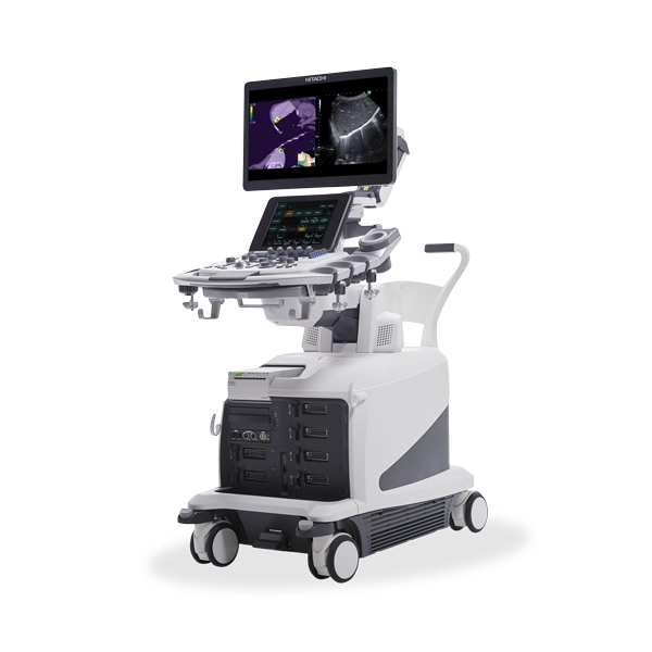
Arietta 850
The next evolution in ultrasound designed for high expectations to answer the ever-increasing demands of medical professionals.
The ARIETTA 850 was designed to address the ever-increasing clinical and operational demands of today’s Radiology suite. It pairs advanced hardware with new and innovative image acquisition techniques to deliver crisp, high-resolution imaging. Efficiency solutions like Protocol Assistant and a new annotation package enable it to be tailored to each individual user’s workflow needs. The ARIETTA 850 expands the overall utility of ultrasound as a modality through optional image fusion, Elastography, and CMUT probe capabilities.
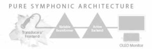
The ARIETTA 850 produces exceptionally clear images powered by the Pure Symphonic Imaging precision technologies, which ensures that only the most relevant data is used to optimize the advanced imaging capabilities of the ARIETTA 850.
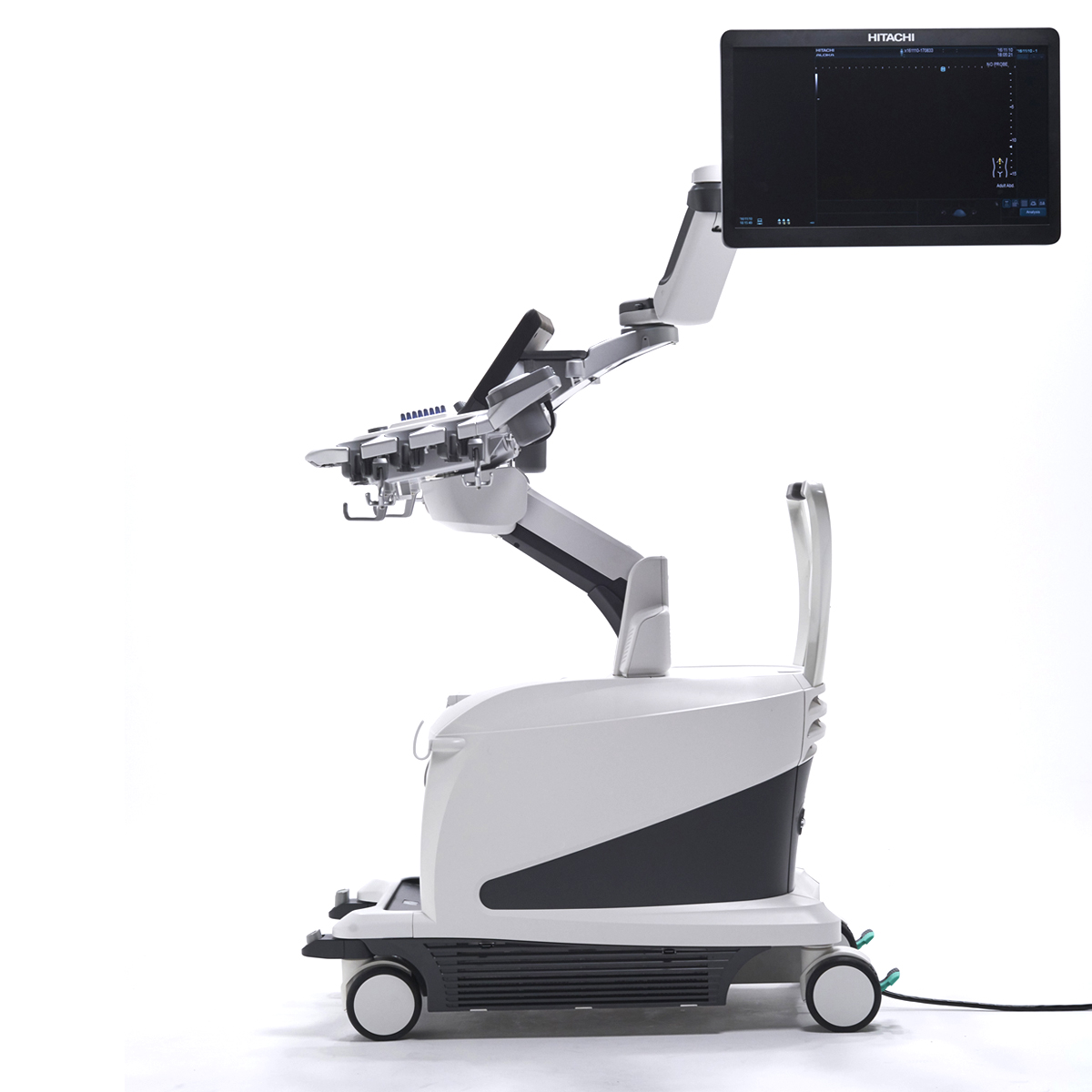
The ARIETTA 850 is designed to help facilitate the workflow while offering an ergonomic work environment to safeguard users from the fatigue and long-term injury that can result from repetitive scanning, allowing more efficiency on the part of the sonographers.
- Radiology and general imaging
- Vascular
- Women’s health
- Cardiovascular
- Surgery
- Oncology brachytherapy
- Point of care (POCUS)
Advanced applications:
- Combinational elastography (SWE, Strain)
- Multi-modality image fusion with real-time virtual sonography (RVS)
- Contrast harmonic imaging
- Fetal 3D/4D imaging
- Advanced cardiac
Through extensive research and user feedback, Hitachi developed tools for workflow enhancement while incorporating advanced workflow features into the ARIETTA 850 series to include:
Ergonomics
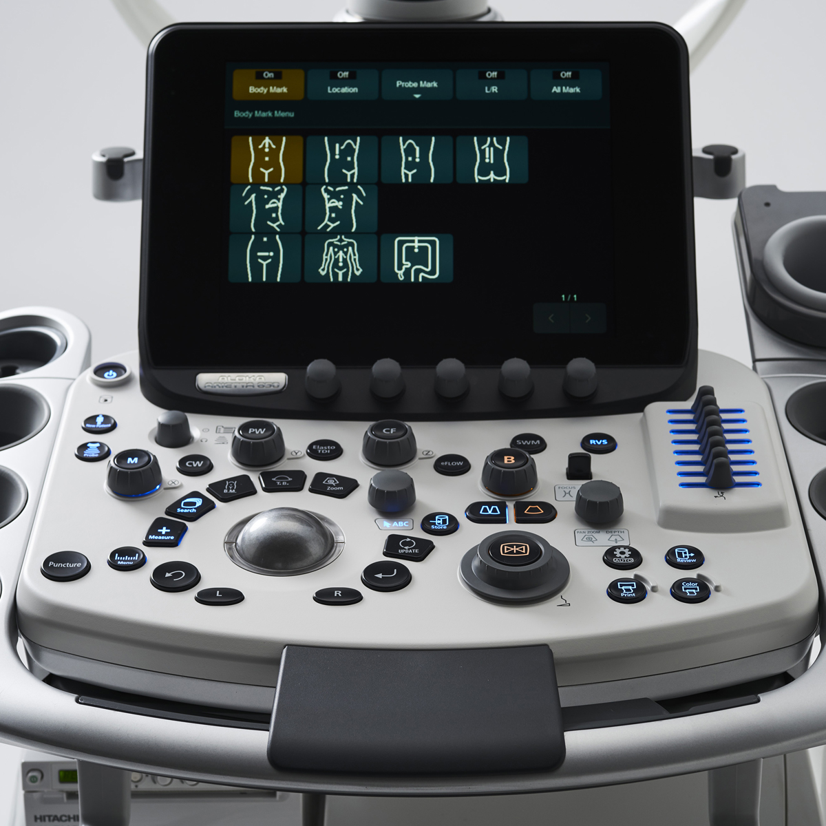
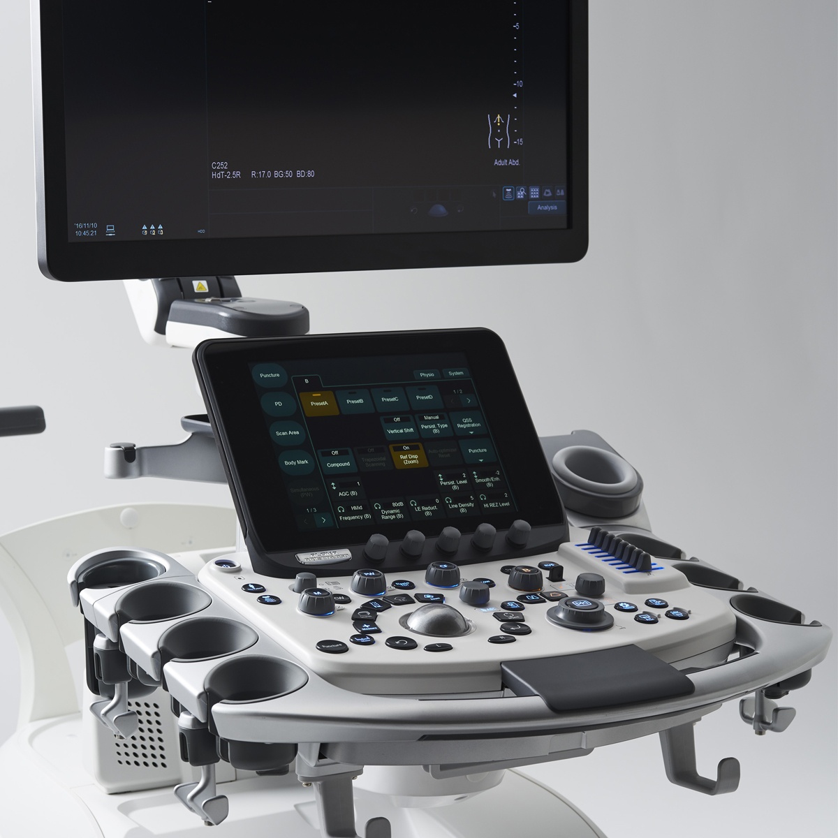
The ARIETTA 850 is engineered to deliver a fast and safe scanning experience. For example, its flexible monitor arm allows front-to-back movement during the exam, independent of positioning. In addition, the intuitive and programmable 5-switch console can simplify advanced functions, measurements and analysis. It is also possible to easily move the console up and down just by pressing the pedal.
OLED Monitors (optional)
The 850’s 22-inch display uses an OLED monitor to deliver exceptional image fidelity. The self-luminous displays true black so a previously unattainable contrast resolution can be achieved, offering a significant improvement in the diagnostic quality of the display.
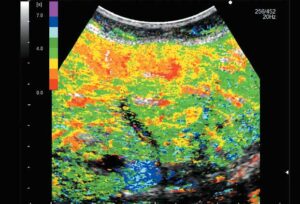
Detective Flow Imaging (DFI)
The new imaging technology for visualization of low velocity blood flow below the previous detection threshold. The unique algorithm displays fine blood flow with greater resolution and sensitivity.
Protocol Assistant
Protocol Assistant can bring standardized scanning protocols to any department in a way that is efficient and reproducible. The system learns how each user wants to perform individual studies and anticipates the user’s next step, providing the correct annotation, system parameters, and measurement tools automatically as each new image is acquired.
Automated Measurements
The ARIETTA 850 automates complex, repetitive measurement routines to simplify time-intensive processes like auto carotid IMT, auto nuchal translucency, auto FHR, Estimated Fetal Weight measurements, optimal frame selection for Elastography interpretation, and placement of Strain Ratio regions of interest.
Adjustable Panel Height
The panel can reach a height of 170 cm and can be lowered to 70 cm in its lowest position, allowing the operator to perform lower extremity examinations with the control panel comfortably within reach.
UNIQUE 4G CMUT Probe
While piezoelectric probe technology has seen little change, ARIETTA 850 has however succeeded in this technological leap, supporting the world’s first fully-featured Capacitive Micro-machined Ultrasound Transducer. The SML44 probe, contains thousands of high-sensitivity, wide-bandwidth CMUT cells that are printed onto a silicon substrate using techniques first developed in the semi-conductor industry. The resulting operating bandwidth of 2-22 MHz, enables the probe to perform the work of multiple conventional probes.
eFocusing
eFocusing employs the ARIETTA 850’s advanced beam former to perform real time focusing along the entire depth of the image. The technique avoids the need to set or adjust focal zones, automatically providing optimal focusing from near to far-field.
Active Backend
Powerful computing and image-processing hardware manages the acquired ultrasound signal, performing countless data-enhancement algorithms like Acoustic Noise Reduction and Nearfield Noise Reduction, while maintaining high frame rates.
In addition to a high-performance ultrasound system, Hitachi has introduced advanced applications to create cost effective uses for this modality to include:
Combinational Elastography (SWE, Strain)
Ultrasound Elastography has been shown to be an accurate non-invasive method of assessing the progression of liver disease by depicting the level of fibrotic change. In the ARIETTA 850, Hitachi has combined the two methods of Elastography (strain and Shear Wave) into a single liver Elastography exam. This fast examination can produce the quantitative data provided by Shear Wave Elastography (SWE) while also exploiting Strain Elastography’s unique advantages. The combination provides a comprehensive picture of liver health, including indices that reflect:
- Overall Fibrotic Progression
- Extent of fatty replacement
- Presence of Inflammation
Dual Gate Doppler
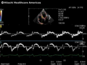
Enables observation of Doppler waveforms from two separate locations during the same heart cycle. Measurements such as the E/e’ ratio can be measured, eliminating beat-to-beat variation.
Multi-modality Image Fusion with Real-time Virtual Sonography (RVS)
Hitachi continues to expand its RVS fusion capabilities with a new suite of visualization and efficiency tools to enhance its ability support a variety of interventional procedures.
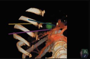
- 3D Sim-Navigator – Provides simulation of single or multiple needle paths during navigation to a target. The positional relationship between the marked target and needle paths can be assessed in real time using the 3D body mark reconstructed from the virtual CT volume data and an additional C-plane display that is orthogonal to the needle path.
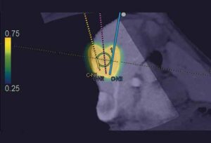
- E-Field Simulator – Designed to streamline RFA procedures, E-field Simulator superimposes a colour map onto the CT image that simulates the estimated distribution of the electric field (E-field) based on the position of multiple electrodes during RFA treatment. The simulation provides instant visual feedback to understand the potential effects of needle positioning.
- Body Motion Tracking – Working in concert with the omniTRAXTM Active Patient Tracker from CIVCOTM, this feature enables instant registration between the live ultrasound image and previously acquired volumes. It also corrects in real time for patient motion during scanning.
- Needle Tracking – Using data from the RVS sensor and the VirtuTRAXTM biopsy bracket from CIVCOTM allows real-time tracking of needle placement and automatically correcting for needle flexion.
- Contrast Harmonic Imaging – With multiple acquisition modes and analytical tools like Inflow Time Mapping, which displays a colour-coded graphical representation of enhancement and wash-out times, the ARIETTA 850 positions users to increase utilization of a technique that has been adopted throughout the rest of the world.
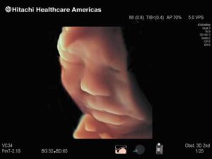
- Fetal 3D/4D Imaging – Add important clinical information to your ultrasound image with our empowered 3D/4D package; while your patients enjoy photo-realistic images that help them bond with their unborn child.
Watch this video:
- Ergonomic work environment to safeguard users from the fatigue and long-term injury that can result from repetitive scanning
- Complete workflow solution
- Focused on patient’s comfort
- Auto optimizer on each mode
- Specific preset parameters that give you immediate high quality images
- Customizable
- Widest range of transducers available on the market
Hitachi Healthcare Americas delivers best in class medical imaging technologies for healthcare providers. Hitachi’s MRI, CT and Ultrasound along with Agfa HealthCare’s Digital Radiology provide speed, comfort and quality for both physicians and patients and play an important role in the diagnosis and treatment of disease while driving social innovation into healthcare. Hitachi’s VidiStar image and reporting platform enables healthcare professionals to create value-based reports leveraging a cloud-based image management and analytics platform for improved communication across the healthcare organization. Physicians can grow their business into pediatrics, orthopedics and other unique patient populations while exploring new areas to compete.
With an optimized and patient centric approach, healthcare providers can deliver strong value into their communities and Hitachi will be there to support them. Our customer first philosophy compels us to make customer support one of the most important things we do. Innovating Healthcare, Embracing the Future.
Request a demo today!
Our innovative Medical Imaging Equipment Solutions and Health IT Solutions are built to support your practice with the latest advancements in medical imaging. Experience how our solutions can enhance diagnostic accuracy, streamline workflows, and improve patient care. Click to request a demo and discover the possibilities firsthand.